Instruments
Details regarding instruments excluded from autonomous operation, as well as the services provided for these instruments, can be found under the Services section.
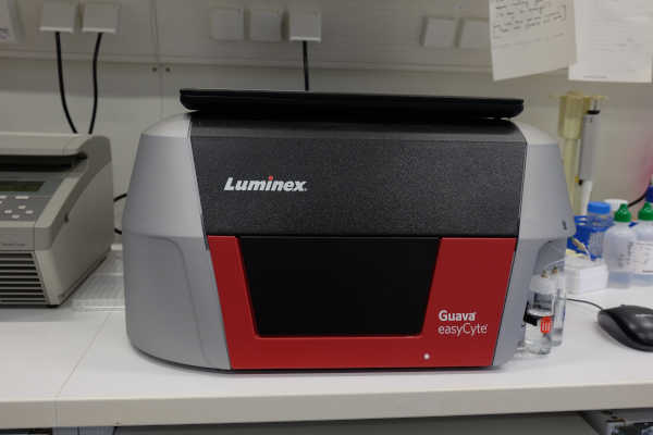
Luminex™ Guava® easyCyte™ Flow Cytometer
The Guava easyCyte is a compact and user-friendly flow cytometer that enables accurate absolute cell counts without the need for reference beads. Utilizing microcapillary flow technology and a precision pump, it operates without sheath fluid, making it ideal for small sample volumes and high-throughput analysis in 96-well plates.
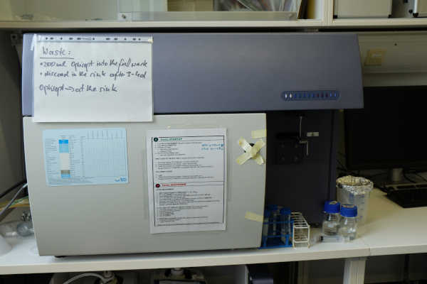
BD® FACSCanto™ II – Flow Cytometer
The BD FACSCanto™ II is a high-performance flow cytometer designed for the simultaneous multiparameter analysis of cell populations. It is equipped with two laser lines—488 nm (blue) and 633 nm (red)—and allows for the detection of up to nine parameters, including forward scatter (FSC), side scatter (SSC), and seven fluorescence channels.
The optical configuration includes the following standard filter sets:
Blue laser (488 nm):
530/30 nm (FITC, GFP)
585/42 nm (PE)
670 LP (PerCP, PerCP-Cy5.5)
Red laser (633 nm):
660/20 nm (APC)
780/60 nm (APC-Cy7)
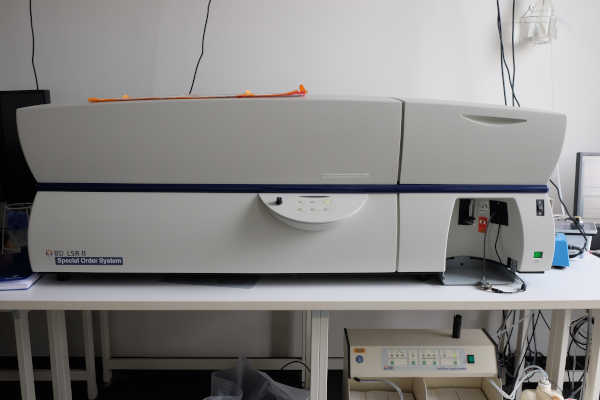
BD® LSR™ II – Flow Cytometer
The BD LSR™ II is a versatile, research-grade flow cytometer designed for high-dimensional multiparameter analysis. It is equipped with three laser lines—405 nm (violet), 488 nm (blue), and 633 nm (red)—and supports the simultaneous detection of up to 17 parameters, including forward scatter (FSC), side scatter (SSC), and 15 fluorescence channels.
The standard optical configuration includes the following filter sets:
Violet laser (405 nm):
450/50 nm (e.g. Pacific Blue)
525/50 nm (e.g. AmCyan)
610/20 nm (e.g. BV605)
710/50 nm (e.g. BV711)
Blue laser (488 nm):
530/30 nm (FITC)
576/26 nm (PE)
695/40 nm (PerCP-Cy5.5)
780/60 nm (PE-Cy7)
Red laser (633 nm):
660/20 nm (APC)
710/50 nm (APC-R700)
780/60 nm (APC-Cy7)
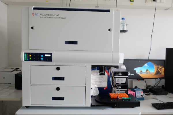
BD® FACSSymphony™ A5 – Flow Cytometer
The BD FACSymphony™ is a state-of-the-art flow cytometry platform designed for high-parameter, high-throughput single-cell analysis. It enables the simultaneous acquisition of an exceptionally high number of parameters per event, offering both high resolution and high acquisition speed. This allows for the detailed characterization of complex cell populations in large sample cohorts.
The system is equipped with a modular laser configuration and can be customized to include up to 5 lasers, such as:
355 nm (UV)
405 nm (violet)
488 nm (blue)
561 nm (yellow-green)
640 nm (red)
Depending on the specific configuration, the FACSymphony can support 30+ fluorescence parameters, making it one of the most advanced analyzers available for multicolor flow cytometry.
An integrated High Throughput Sampler (HTS) is available for automated sample acquisition from 96-well plates, significantly increasing throughput and experimental efficiency.
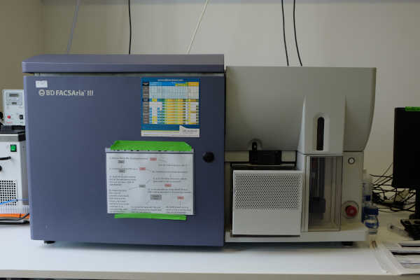
BD® FACSAria™ III – Flow Cytometer Cell Sorter
The BD FACSAria™ III is a high-speed, high-precision cell sorter designed for the simultaneous isolation of multiple cell populations. It supports sorting into tubes as well as 96- and 384-well plates, enabling flexible workflows for various experimental needs.
The system allows for the simultaneous sorting of up to four distinct populations, using customizable gating strategies and high-speed droplet deflection. Its fixed optical alignment with a cuvette-based flow cell ensures consistent signal detection, high sensitivity, and reduced setup complexity.
The BD FACSAria III can be configured with up to six lasers, allowing for the detection of a wide range of fluorochromes. A common configuration includes the following lasers:
405 nm (Violet)
488 nm (Blue)
561 nm (Yellow-Green)
633 nm (Red)
This configuration enables the detection of up to 18 colors simultaneously, providing flexibility for complex multicolor experiments
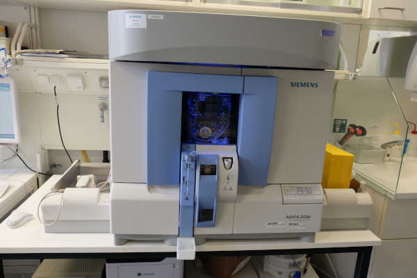
ADVIA® 2120i Hematology System
The ADVIA 2120i is a high-throughput, fully automated hematology analyzer designed for comprehensive blood cell analysis. It provides precise and reliable results using advanced flow cytometry and cytochemical staining methods. With minimal sample preparation and integrated automation, it enables efficient processing of up to 120 samples per hour, making it ideal for routine and high-volume clinical laboratories.
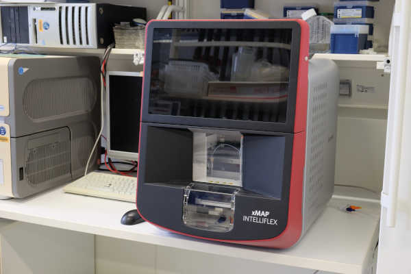
Luminex™ xMAP™ INTELLIFLEX® System
The xMAP INTELLIFLEX® is a next-generation multiplexing platform developed by Luminex Corporation. It enables the simultaneous detection and quantification of multiple analytes—such as proteins, nucleic acids, or other biomolecules—in a single sample. Utilizing advanced flow cytometry and bead-based technology, the system offers high-throughput analysis with enhanced sensitivity and flexibility, making it ideal for applications in research, clinical diagnostics, and drug development.
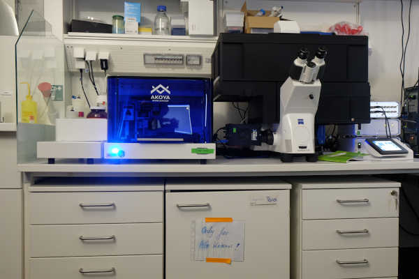
Akoya PhenoCycler™ (formerly CODEX®) – High-Parameter Spatial Biology Platform
The PhenoCycler™ (formerly known as CODEX® – CO-Detection by indEXing) is an advanced multiplexed imaging system developed by Akoya Biosciences. It enables high-throughput, spatially resolved detection of dozens of protein markers in a single tissue section while preserving cellular morphology and spatial context. Using DNA-barcoded antibodies and iterative fluorescence imaging, the PhenoCycler™ system provides powerful insights into tissue architecture and cellular interactions—making it a key tool in immuno-oncology, neuroscience, and translational research.
Acknowledgements
The Core Facility thank the German Research Foundation for continuous support.